![]()
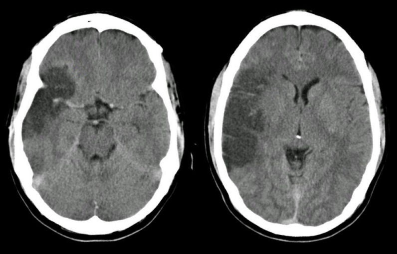
| Superior Division Middle Cerebral Artery (MCA) Infarction:
Axial
CT scans 3 days after presentation.
The CT scans show a hypodensity primarily affecting the territory of the superior division of the right middle cerebral artery, involving the right frontal lobe above the
Sylvian
fissure. The internal carotid artery terminates in a larger MCA and smaller anterior cerebral artery. The MCA runs horizontally to the Sylvian fissure, giving off the lenticulostriate vessels. These small perforating vessels supply the basal ganglia and internal capsule. The MCA then typically bifurcates into a superior and inferior division. The superior division supplies the lateral frontal and superior parietal lobes, whereas the inferior division predominantly supplies the lateral temporal and inferior parietal lobes. The superior division of the MCA is one of the most common locations for an embolic stroke, either from a proximal arterial source or from the heart. Complete infarctions typically result in a contralateral hemiparesis affecting the lower face and upper extremity more than the leg; contralateral hemisensory loss with a similar distribution; contralateral visual field deficit predominantly affecting the lower fields; and often a gaze preference to the ipsilateral side. With an infarct in the dominant hemisphere, there is often an associated expressive aphasia; with a non-dominant infarct, there is often a neglect syndrome. |
Revised
11/22/06
Copyrighted 2006. David C Preston