![]()
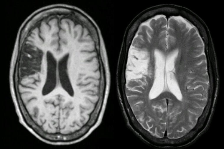
| Superior Division Middle Cerebral Artery (MCA) Infarction: (Left) T1-weighted axial MRI; (Right) T2-weighted axial MRI. Note the well demarcated lesion in the distribution of the superior division of the right middle cerebral artery, involving the right frontal lobe above the Sylvian fissure. Note that the stroke is dark on the T1-weighted scan and bright on the T2-weighted scan, similar to CSF. In the chronic state, strokes result in encephalomalacia, wherein the infarcted area of brain undergoes necrosis, leading to an essentially cystic space. Also note that the ventricle on the right is slightly larger than the one on the contralateral side (i.e., hydrocephalus, ex vacuo). This is the picture of a remote superior division MCA stroke. In this case, the remote stroke was the etiology of the patient's seizures. Indeed, in older individuals, remote cortical stroke is the most common cause of new onset seizures. |
Revised
11/27/06
Copyrighted 2006. David C Preston