|
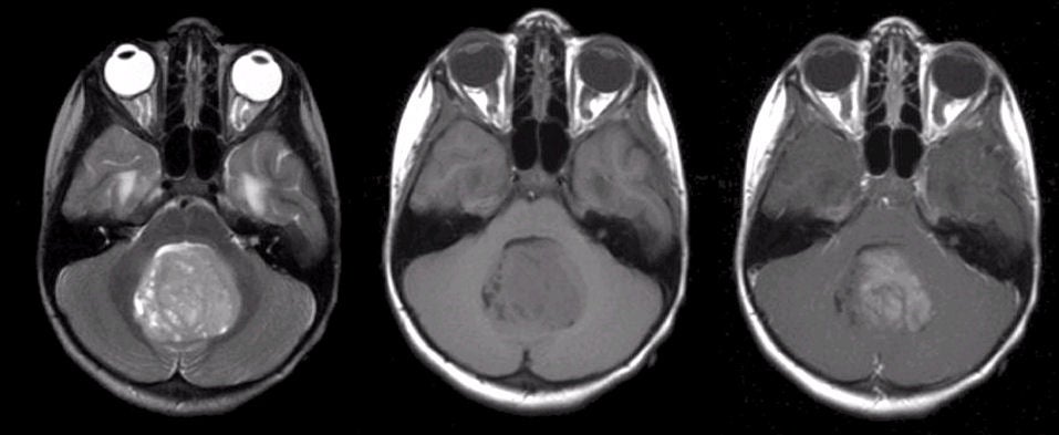
A 4 year-old boy presented with headaches, vomiting and an unsteady gait. |
![]()
![]()
![]()
| Medulloblastoma. (Left) T2-weighted axial
MRI; (Middle)
T1-weighted axial MRI; (Right) T1-weighted with gadolinium axial MRI. Note the large enhancing mass invading the roof of the fourth ventricle.
Compression of the fourth ventricle resulted in non-communicating
hydrocephalus. Surgical resection demonstrated the mass to be a medulloblastoma. Medulloblastoma most often occurs in children and is responsible for approximately 30% of intracranial pediatric tumors. It is one of the primitive neuroectodermal tumors (PNET). The tumor usually arises in the midline of the roof of the fourth ventricle and may spread locally as well as to other parts of the neuraxis, especially the meninges and spinal cord, the latter known as "dropped mets." |
Revised
11/27/06.
Copyrighted 2006. David C Preston