![]()
![]()
![]()
![]()
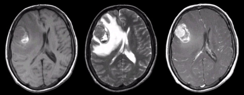
| Metastatic Brain Tumor (Melanoma):
Axial MRI images: (Left) T1-weighted; (Middle) T2-weighted; (Right) T1-weighted with gadolinium.
Note the large mass in the right frontal lobe that enhances with
gadolinium (right image). On the T1-weighted image (left image), note the
bright signal
within the mass. On the T2-weighted image (middle image), there is a dark rim surrounding a
dark signal. This is the MRI picture of subacute blood (intracellular methemoglobin).
Melanoma is one of the metastatic tumors that commonly bleeds.
Metastatic disease from primary tumors elsewhere in the body account
for approximately 50% of all brain tumors. Metastases to the brain
are nearly always via the blood stream. They are typically found at
the junctions between the gray and white matter, which are highly
vascular. Metastatic lesions commonly present with focal or focal to
generalized seizures or slowly progressive neurological deficits.
When the lesions become very large, signs and symptoms of increased
intracranial pressure develop (i.e., headache, lethargy, nausea and
vomiting). The most common primary tumors that metastasize to the
brain are lung and breast. Other tumors may also spread to the
brain, including melanoma, lymphoma, GI, and GU cancers. In some
cases, it is the metastatic lesion in the brain, and not the primary
tumor, that brings the patient to medical attention. |
Revised
11/29/06.
Copyrighted 2006. David C Preston