|
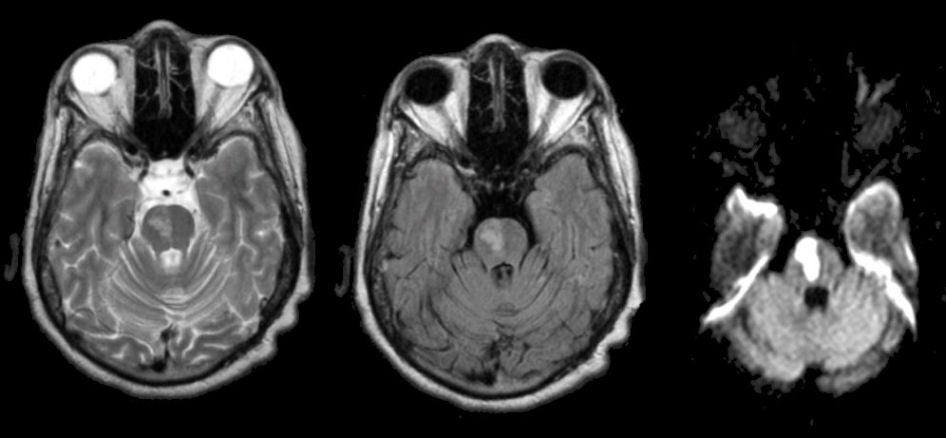
A 72 year-old woman with diabetes developed dysarthria and a slight left hemiparesis. |
![]()
| Pontine Infarction: (Left) T2 weighted-axial MRI; (Middle) Flair axial MRI; (Right) Diffusion-weighted MRI. Note the bright signal in the right pons. On the diffusion-weighted image, the lesion is bright, denoting that it is acute. This infarct is in the distribution of one perforating branch of the basilar artery. This lesion is usually caused by the occlusion of one perforating basilar branch, due to lipohyalinosis, which occurs as a result of aging, diabetes and hypertension. Occasionally these lesions are associated with intrinsic disease of the basilar artery or an embolus to the basilar artery. |
Revised
11/23/06
Copyrighted 2006. David C Preston