|
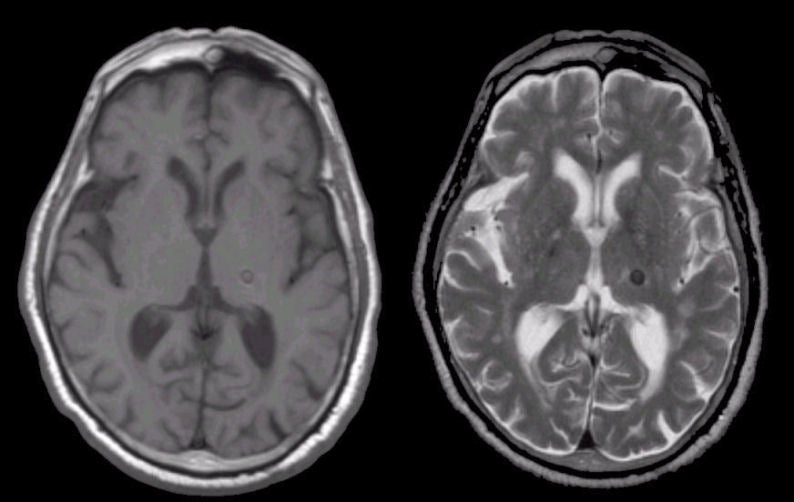
A 47 year-old woman presented with headaches. |
![]()
![]()
| Remote Intracerebral Hemorrhage: (Left) T1-weighted axial MRI; (Right) T2-weighted axial weighted MRI. Note the lesion that is dark on both T1- and T2-weighted images. This picture is consistent with hemosiderin, the residual from old blood. In this case, the old hemorrhage was caused by a cavernous angioma. To learn more, review the powerpoint slide show, Blood on MRI: Time-dependent Changes. |
Revised
11/29/06.
Copyrighted 2006. David C Preston.