|
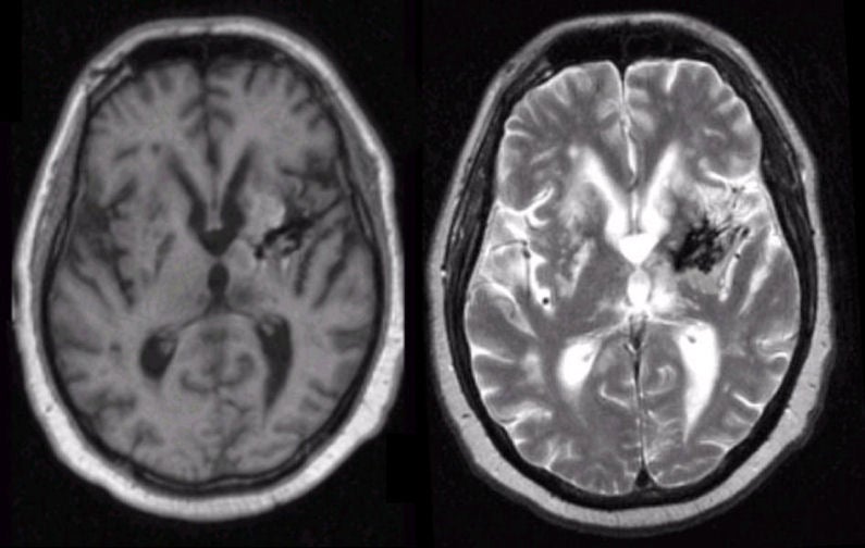
A 77 year-old man presented with focal seizures affecting the right side of the body. There was a history of an intracerebral hemorrhage four years earlier. |
![]()
![]()
| Remote Intracerebral Hemorrhage: (Left) T1-weighted axial MRI; (Right) T2-weighted axial MRI. Note the lesion in the left basal ganglia that is dark on both T1- and T2-weighted images. This picture is consistent with hemosiderin, the residual from old blood. In this case, the hemorrhage was due to hypertension. To learn more, review the powerpoint slide show, Blood on MRI: Time-dependent Changes. |
Revised
11/29/06.
Copyrighted 2006. David C Preston.