![]()
| Spinal Arteriovenous Malformation (AVM):
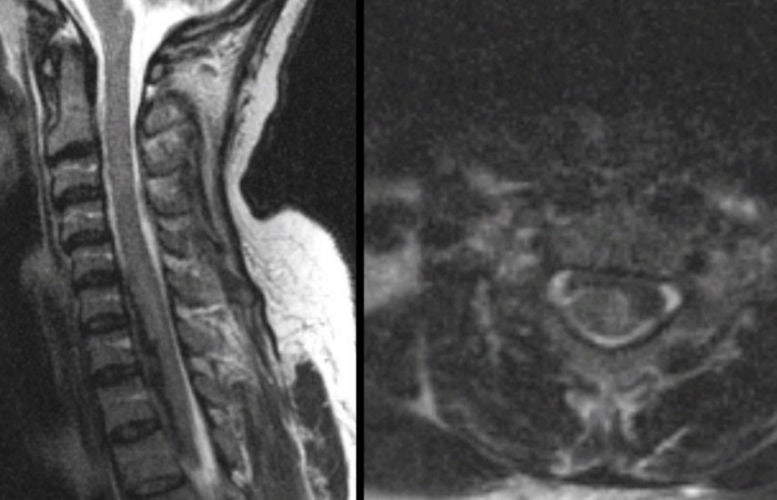
(Left) T2-weighted sagittal MRI of the cervical spine; (Right) T2-weighted axial
MRI of the cervical spine. Note the
lesion that extends from C6 - T2, which is dark on T2-weighted
images. It likely
represents deoxyhemoglobin or intracellular methemoglobin. On the axial image,
note that the lesion appears to be subdural in location. This is a subacute
subdural
hematoma which occurred from a ruptured spinal AVM. Arteriovenous malformations
are a congenital abnormality of blood vessels, consisting of a
tangle of abnormal vessels supplied by arterial feeders and often
drained by large dilated veins. Spinal cord vascular malformations may be acquired or congenital. They are malformations of blood vessels in or around the spinal cord, and may take on several forms, including arteriovenous malformations, dural arteriovenous fistulas, hemangiomas, cavernous angiomas, and aneurysms. The clinical presentation depends on whether the bleed (acute presentation) creates a vascular steal phenomena, resulting in chronic spinal cord ischemia, or enlarges and creates a defacto mass effect on the spinal cord, resulting in spinal cord compression. They are potentially treatable by neurosurgical decompression or endovascular procedures. |
Revised
11/18/06
Copyrighted 2006. David C Preston