|
A 72 year-old woman presented with the acute onset of a left hemiplegia affecting the face, arm and leg, associated with a neglect syndrome and right eye deviation. After the initial CT showed no evidence of hemorrhage, IV tPA was administered followed by intra-arterial tPA. Two hours later, she was in a coma with bilateral Babinksi signs. |
![]()
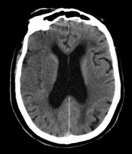
| Top of the Carotid Occlusion:
Axial CT scan, Pre-tPA. Other than some periventricular white matter
disease and atrophy, this scan is unremarkable.
However, in the Post-tPA scan, there is a new massive intracranial
hemorrhage in the region of the right basal ganglia with mass
effect, midline shift and extension into the ventricles. This is the most dreaded complication of tPA - intracranial hemorrhage. It occurs in 6% of patients treated with tPA, and half of those hemorrhages are fatal. In this case, the patient was pronounced brain dead six hours later. Although this patient had a PComm aneurysm, it was probably incidental, with the hemorrhage occurring as a complication of the tPA. |
Revised
11/15/06
Copyrighted 2006. David C Preston