|
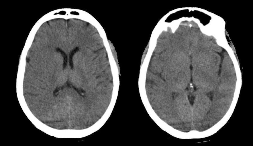
A 63 year-old woman with atrial fibrillation presented with the acute onset of a left hemiplegia and neglect syndrome. After the CT showed no evidence of hemorrhage, IV tPA was administered followed by intra-arterial tPA. The next day, she still had a dense left hemiplegia with her eyes deviated to the right. |
![]()
![]()
![]()
| Middle Cerebral Artery Occlusion: Axial CT scans (Initial Scans). These scans are normal except for the slight loss of gray-white differentiation over the right hemisphere, with subtle effacement of the sulci. Such findings on a scan as early as this suggest an evolving very large ischemic infarct. Despite successful recanalization with tPA, the patient remained unchanged 1 day later, with a major stroke. The stroke is well seen on the subsequent CT scans, and is so massive that it is associated with mass effect and midline shift. Note also that there is some hemorrhagic conversion on the second set of scans. |
Revised
11/29/06
Copyrighted 2006. David C Preston