|
A 47 year-old woman presented with headaches. She had an episode of transient right sided numbness several years earlier. |

![]()
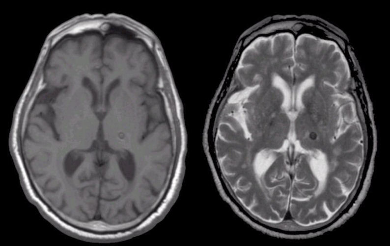
| Cavernous Angioma:
(Left) T1-weighted axial MRI;
(Right) T2-weighted axial MRI. Note the lesion which is dark on both T1- and
T2-weighted images. The lesion is in the left thalamus and is likely
the cause of the patient's episode of right sided numbness years
ago. This pattern of being dark on both T1- and T2-weighted MRI is
characteristic of hemosiderin, the residual of old blood.
This lesion is a cavernous angioma. Cavernous angiomas are a type of intracranial vascular malformation. They typically occur in the deep white matter, basal ganglia, or frontal or temporal lobes, and less commonly in the pons and cerebellum. With the advent of MRI, congenital cavernous angiomas are more often recognized as a source of intracerebral hemorrhage. They are often asymptomatic unless they bleed, when patients present with seizures, focal neurological deficits, or signs and symptoms of increased intracranial pressure, similar to intracerebral hemorrhages from other causes. |
Revised
11/30/06
Copyrighted 2006. David C Preston