|
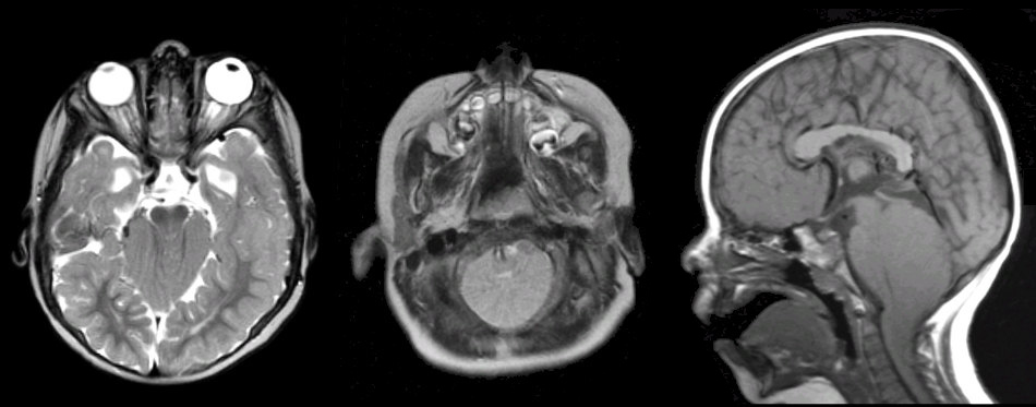
A 9 month-old boy presented with an enlarging head and a delay in motor milestones. |

SHOW THE FINDINGS IN THE CHIARI 2 MALFORMATION
![]()
![]()
![]()
![]()
| Chiari Malformation Type II:
(Left and Middle) T2-weighted
axial MRIs; (Right) T1-weighted sagittal MRI. The axial scans show
several abnormalities, including descent of the cerebellar tonsils, an abnormal midbrain with a malformed "beaked" tectum (fusion of the colliculi),
and enlarged ventricles (hydrocephalus). The sagittal scan shows several abnormalities,
including
descent of the cerebellar tonsils, a "kinked" medulla, and a very
tight posterior fossa.
This malformation is know as a Chiari malformation, type II. It is
quite complex and associated with more serious consequences than the
type I malformation. The type II malformation affects the entire
hindbrain, dura and bone, as well as the spinal cord, the latter
presenting as spina bifida and a meningomyelocele. Among the
abnormalities included in this malformation are aqueductal stenosis,
hydrocephalus, tonsillar herniation, a "kinked" medulla, an
elongated 4th ventricle, fused colliculi resulting in a "beaked"
tectum of the midbrain, partial agenesis of the corpus callosum,
syringomyelia, and a dysplastic skull. |
Revised
11/22/06
Copyrighted 2006. David C Preston