|
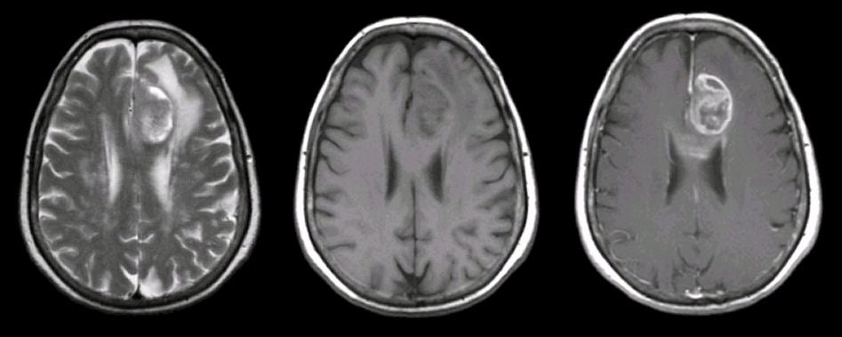
A 47 year-old woman presented with progressive spasticity of the right lower extremity associated with headaches and a depressed affect. |
![]()
![]()
![]()
| Glioblastoma Multiforme (Frontal Lobe).
(Left) T2-weighted axial MRI; (Middle)
T1-weighted axial MRI; (Right) T1-weighted with gadolinium axial MRI. Note the large, well demarcated mass in the left frontal lobe that enhances with
gadolinium (GAD). There is also
a cystic component to the tumor. Surgical biopsy confirmed the diagnosis of
malignant glioblastoma. Glioblastoma multiforme (GBM), also referred to
as a Grade IV astrocytoma, is the most common type of primary brain
tumor. It is a malignant tumor that carries a very poor prognosis,
and typically results in death in 2 years. On CT and MRI imaging,
the tumor is often large, irregular and infiltrative, and located in
the white matter with surrounding edema. Histologically, the tumor
is highly cellular and anaplastic with necrosis. Associated
hemorrhage is not uncommon. Clinically, patients present with slowly progressive focal neurological signs, and signs of increased intracranial pressure (i.e., headache, nausea, and vomiting). Seizures may be an initial presentation or may occur later in the course. |
Revised
11/30/06.
Copyrighted 2006. David C Preston