 |
|
|
|
|
|
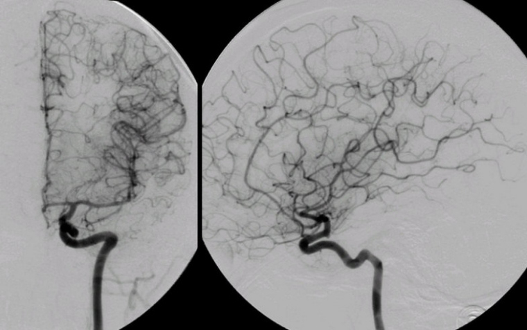
Internal Carotid Angiogram: (Left) AP View; (Right) Lateral View The Internal Carotid Artery (ICA) is commonly divided into segments: (1) The Cervical segment runs from above the carotid bulb through the neck to the base of the skull; (2) the Petrous segment runs from the base of the skull through the petrous bone; (3) the Cavernous segment runs through the cavernous sinus (note the prominent bends); and (4) the Supraclinoid segment runs above the clinoid process through the dura into the subarachnoid space; several important branches arise from the supraclinoid carotid, among them the ophthalmic, posterior communicating, and anterior choroidal arteries. The Middle Cerebral Artery (MCA) is commonly divided into four numbered segments: (1) the M1 segment runs horizontally from the origin of the MCA to the Sylvian fissure; (2) the M2 segments runs vertically adjacent to the insula; (3) the M3 segments runs adjacent to the operculum from the insula to the surface of the Sylvian fissure; and (4) the M4 segments run from the surface of the Sylvian fissure to over the cortex. Typically, the horizontal segment (M1) of the MCA bifurcates into a superior and inferior trunk. Other common patterns include a trifurcation or a prominent anterior temporal branch arising from the M1 segment (as seen in the case above). The Anterior Cerebral Artery (ACA) is commonly divided into three numbered segments: (1) the A1 segment runs horizontally and medially from the origin of the ACA to where the anterior communicating artery arises; (2) the A2 segment runs vertically from the where the anterior communicating artery arises to genu of the corpus callosum; and (3) the A3 segments run to supply the cortex. |