|
A 44 year-old woman presented with headaches, worse when lying down. Her neurological examination was normal. |

![]()
![]()
![]()
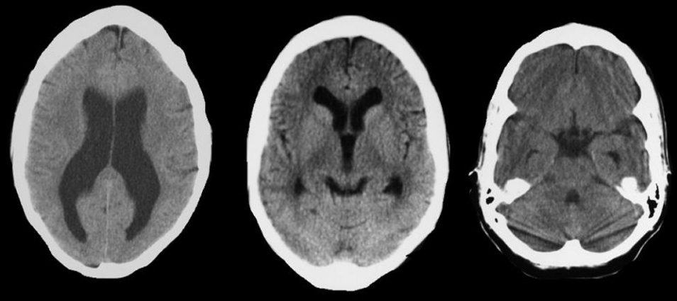
| Non-Communicating Hydrocephalus:
Axial CT scans. Note the
prominent enlargement of the lateral and third ventricles in the setting of a normal sized
fourth ventricle. This pattern is one of non-communicating
(obstructive) hydrocephalus, which occurs from impaired drainage through the cerebral aqueduct
which connects the third and fourth ventricles. This picture
differs from communicating hydrocephalus wherein all the ventricles
are enlarged. Note also that the frontal horns are enlarged -
they extend beyond the head of the caudate nucleus and form somewhat
of a picture of "mickey mouse" ears. Note also that the
sulci are relatively effaced (i.e. small), which excludes the possibility that the
enlarged
ventricles are simply due to cerebral atrophy (i.e., hydrocephalus ex vacuo). The
amount of ventricular dilatation seen here is mild, and likely
represents a chronic compensated state. In a compensated state, the
ventricles enlarge to a point whereby the increased pressure results
in CSF absorption being equal to CSF production. In this situation,
patients may have little or no symptoms. Hydrocephalus is recognized as enlarged ventricles out of proportion to the amount of cerebral atrophy. Non-communicating (obstructive) hydrocephalus occurs when the ventricular system is not in continuity with the subarachnoid space. Most often, the site of the blockage in non-communicating hydrocephalus is at the cerebral aqueduct, but rarely can occur at the foramen of Monro, the third ventricle, or the outlet of the fourth ventricle. Non-communicating hydrocephalus is an important disorder to recognize as it is potentially treatable by shunting. |
Revised
11/29/06
Copyrighted 2006. David C Preston