|
A 23 year-old woman presented with headaches and difficulty with gait. Examination was notable for bilateral papilledema. |
![]()
![]()
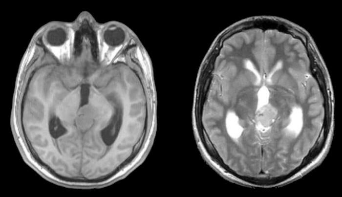
| Pinealoma:
(Left) T1-weighted axial MRI; (Right) T2-weighted axial MRI. Note the lesion located in the posterior third ventricle,
associated with hydrocephalus. Surgical excision showed that the
lesion was a pinealoma. Tumors that arise from cells in the pineal gland are known as pinealomas. As the pineal gland is located in the posterior third ventricle, pineal tumors often present with non-communicating hydrocephalus due to compression of the cerebral aqueduct. In addition, downward pressure may compress the dorsal midbrain, resulting in a Parinaud's syndrome (lid retraction, large pupils that react poorly to light, impaired upgaze, and convergence retraction nystagmus). |
Revised
11/28/06.
Copyrighted 2006. David C Preston