|
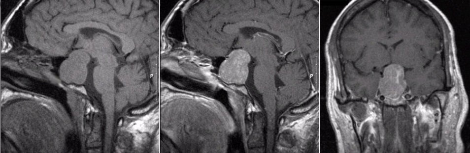
A 44 year-old woman developed progressive headaches. On examination, she was found to have a dense bitemporal hemianopsia. |
![]()
![]()
![]()
| Pituitary Macroadenoma. (Left) T1-weighted
sagittal MRI; (Middle)
T1-weighted with gadolinium sagittal MRI; (Right) T1-weighted with
gadolinium coronal MRI. Note the large enhancing mass in the region of the sella
that is growing up into and displacing the optic chiasm and
hypothalamus. Surgical resection demonstrated a large pituitary
adenoma. By definition, pituitary macroadenomas are benign tumors
of the pituitary gland that are greater than 10 mm in diameter.
Similar to microadenomas, macroadenomas may come to medical
attention due to signs and symptoms of endocrine dysfunction from
excessive hormonal production. However, in contrast to microadenomas,
macroadenomas may result in reduced hormone production of some or
all of the pituitary hormones (panhypopituitarism) as the tumor
grows and compresses the normal pituitary tissue. Macroadenomas may
also result in focal neurological signs and symptoms due to mass
effect as the tumor grows outside of the sella and compresses the
optic chiasm and hypothalamus above. Lesions of the optic chiasm
classically result in a bitemporal hemianopsia. |
Revised
11/27/06.
Copyrighted 2006. David C Preston