|
An 82 year-old man presented with focal seizures affecting the right arm. There was a longstanding history of hypertension and a history of a "stroke" years ago that resulted in right sided weakness that resolved over 6 months. |
![]()
![]()
![]()
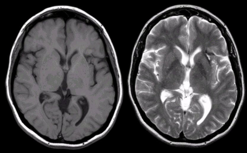
| Remote Intracerebral Hemorrhage: (Left) T1-weighted
axial MRI; (Right) T2-weighted axial MRI. Note that
on the T1-weighted scan, there is an area of hypodensity between the putamen, anterior limb of the internal capsule and insula. The same
area on the T2-weighted scan is also dark, but has a bright center.
This is the characteristic picture of remote hemorrhage with hypointense (dark) signal on T1- and T2-weighted scans, representing hemosiderin. The small area of bright signal on the T2-weighted scan is fluid, which sometimes remains as a collapsed cyst following hemorrhage and destruction of brain tissue. The "stroke" years ago was a hemorrhage, perhaps due to his longstanding hypertension. The findings of blood on MRI are complex and depend on timing. To learn more, review the powerpoint slide show, Blood on MRI: Time-dependent Changes. |
Revised
11/11/06.
Copyrighted 2006. David C Preston.