|
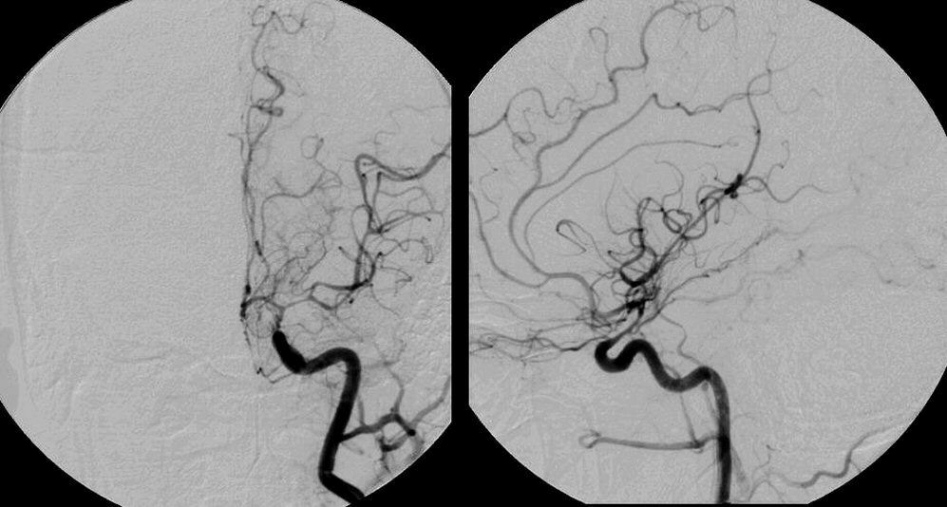
A 44 year-old man presented with a sudden, severe headache, nausea and vomiting. CT scan showed acute subarachnoid hemorrhage. On angiogram, no source of bleeding was seen. Five days later, he developed aphasia and a right hemiparesis. |

![]()
| Vasospasm following Subarachnoid Hemorrhage. Cerebral angiogram,
left internal carotid
artery injection. (Left) AP view; (Right) Lateral view. Note the severe narrowing of the
top of the carotid, middle cerebral, and anterior cerebral arteries. This is the
picture of vasospasm, a known delayed complication of subarachnoid
hemorrhage (SAH). In a small number of cases of subarachnoid
hemorrhage, no bleeding source is identified, despite repeated
angiograms. It is postulated that this may occur from a small
vascular malformation that obliterates itself when it ruptures. ICA = internal carotid artery, MCA = middle
cerebral artery, ACA = anterior cerebral artery. Vasospasm is reported to occur in as many as 70% of patients with SAH and is clinically symptomatic in as many as 30% of patients. It most commonly occurs 4-14 days after the onset of bleeding. If severe enough, vasospasm may lead to progressive ischemia and stroke. In some cases, the acute stroke then results in edema, herniation and death. Vasospasm is typically treated with the calcium channel blocker nimodipine, volume expansion, mild elevation of blood pressure, and in some cases, angioplasty of the involved blood vessels. All of these treatments are better tolerated after the aneurysm has been successfully treated. |
Revised
11/18/06.
Copyrighted 2006. David C Preston