|
A 72 year-old woman presented with the acute onset of a left hemiplegia affecting the face, arm and leg, associated with a neglect syndrome and right eye deviation. After the initial CT showed no evidence of hemorrhage, IV tPA was administered followed by intra-arterial tPA. Two hours later, she was in a coma with bilateral Babinksi signs. |
![]()
![]()
![]()
![]()
![]()
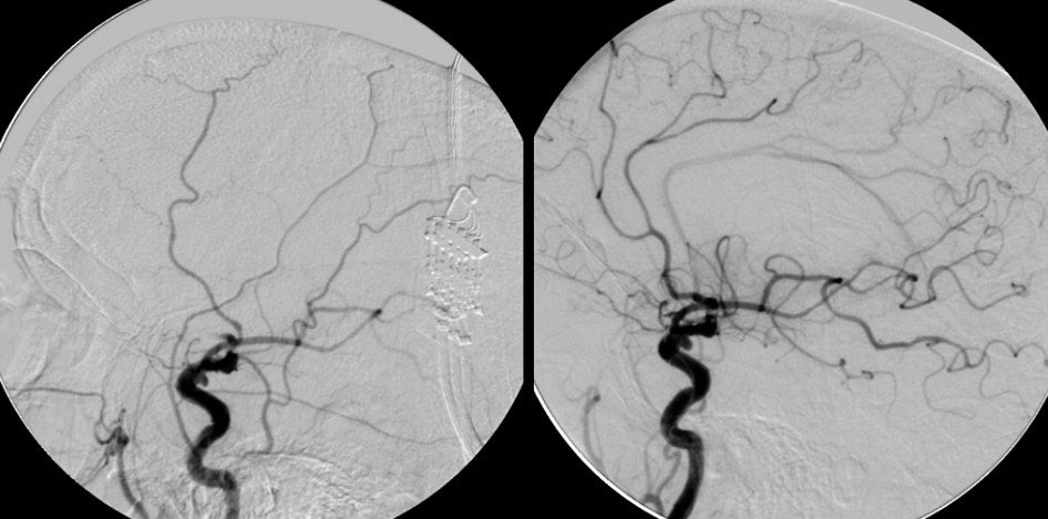
| Top of the Carotid Occlusion: Cerebral angiogram, lateral view, right internal carotid artery (ICA) injection; (Left) Pre-tPA; (Right) Post-tPA. On the Pre-tPA film, note the occlusion at the top of the carotid artery with no filling of the anterior cerebral artery (ACA), and minimal filling of the middle cerebral artery (MCA). Indeed, the vessels above the internal carotid artery are actually branches off the external carotid artery (ECA) supplying the scalp. On the Post-tPA film, the ACA is well seen as are some of the M2 and M3 branches off the MCA. However, also note that there is a small posterior communicating (PComm) artery aneurysm present. |
Revised
11/22/06
Copyrighted 2006. David C Preston