|
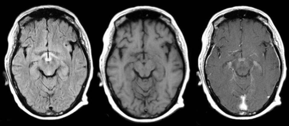
A 50 year-old woman with intractable vomiting developed confusion, poor memory and loss of vision. |

![]()
![]()
![]()
| Wernicke's Encephalopathy:
(Left) Flair axial MRI, (Middle) T1-weighted axial MRI, (Right) T1-weighted with gadolinium
axial MRI.
Note the enhancement of the mammillary bodies on the
post-gadolinium MRI scan (Right) and the prominent signal changes in the hypothalamus and
mammillary bodies on the Flair image
(Left),
changes typical of Wernicke's encephalopathy. Wernicke's encephalopathy results from thiamine deficiency, and constitutes a medical emergency. Left untreated, coma and death may ensue within days. It is seen most often in alcoholics, but also occurs in the setting of poor nutrition from many other causes, including bariatric surgery, morning sickness associated with pregnancy, and intractable vomiting associated with chemotherapy. The classic triad of Wernicke's encephalopathy includes mental state changes, ataxia, and ophthalmoplegia (especially cranial nerve VI). However, all three components may not be present initially. In other cases, Wernicke's encephalopathy may present as seizures, including status epilepticus, visual loss, or coma. The eye findings in Wernicke's encephalopathy include gaze palsies, partial palsies of cranial nerves III, IV, or VI, and nystagmus. This patient recovered completely several days after thiamine supplementation. |
Revised
11/29/06.
Copyrighted 2006. David C Preston