discover
AUDACIOUS OPHTHALMOSCOPE
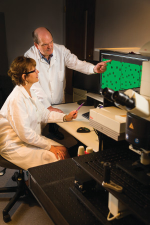 photo: Anthony Gray
Grazyna Palczewska and Krzysztof Palczewski view images captured with the ophthalmoscope their research team is developing that shows age-related degenerative changes in mice.
Krzysztof "Kris" Palczewski wants to look into the eye and see the future. Then, he intends to change it.
The Case Western Reserve professor is creating an ophthalmoscope that can see eye disease developing before noticeable structural changes occur.
Unlike current technologies, which show only the structures within the eye, Palczewski's scope will allow physicians to watch the dance of molecules as they turn light into nerve impulses.
This more precise view should help clinicians predict and treat oncoming diseases, such as age-related macular degeneration, before they have done their greatest damage. Because the device also will test vision, it could enhance researchers' ability to test the efficacy of new treatments.
"Today, if I had a great idea to treat age-related macular degeneration, I would have to wait 50 years to see if this new drug works," said Palczewski, PhD, the John H. Hord Professor and chair of the Department of Pharmacology in the School of Medicine. "If I had this device, which assesses both structure and function quickly, then we shorten the time of testing medications significantly."
With a $3.3 million, five-year grant from the National Eye Institute (NEI), Palczewski and his team hope to take what they learned from building a scope used successfully with mice and build a comparable device for humans. The grant, announced in May, is part of NEI's Audacious Goals Initiative, and one of only five nationwide.
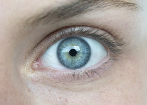
To examine the eye in detail, Palczewski needs to see behind the retina, a light-sensitive layer at the back of the eye that features more than 100 million tiny light-sensing nerve cells called rods and cones. Behind the rods and cones sits a layer of cells responsible for both nourishing the retina and removing accumulating waste. Disease damage first appears in this layer. In vision, it produces molecules that fluoresce in the presence of light, which Palczewski's new scope will image.
Ultraviolet (UV) light was long thought the only way to create images of the fluorescing molecules, but UV light damages tissue. To get around this substantial barrier, Palczewski uses an approach called two-photon excitation, shooting two photons of less-energetic infrared light at a target molecule in rapid succession. The photons arrive so close together—less than a quadrillionth of a second apart—they have the same impact of a single ultraviolet photon.
To focus the image of the fluorescing molecules, Palczewski borrows an idea employed by NASA to correct the blurred vision of the Hubble Space Telescope. As the ophthalmoscope creates the image, 128 tiny mirrors, each capable of independent movement, quickly adjust their positions to eliminate any blurriness.
"This research is on the cutting edge," Palczewski said. "That's why this grant is audacious. We still have a lot to learn in the next two or three years, but I think it will make a major breakthrough."
Palczewski is collaborating with scientists from Harvard University, the University of Pennsylvania, Washington University in St. Louis, Cleveland Clinic and University Hospitals Case Medical Center. The Cleveland-based biotech company Palczewski helped launch, Polgenix, also works on the project. Palczewski and his wife, Grazyna Palczewska, have an equity stake in Polgenix, where he is chief scientific officer and she is director of medical devices.