|
A 32 year-old woman presented with the abrupt onset of a severe headache associated with a mild left hemiplegia. |
![]()
![]()
![]()
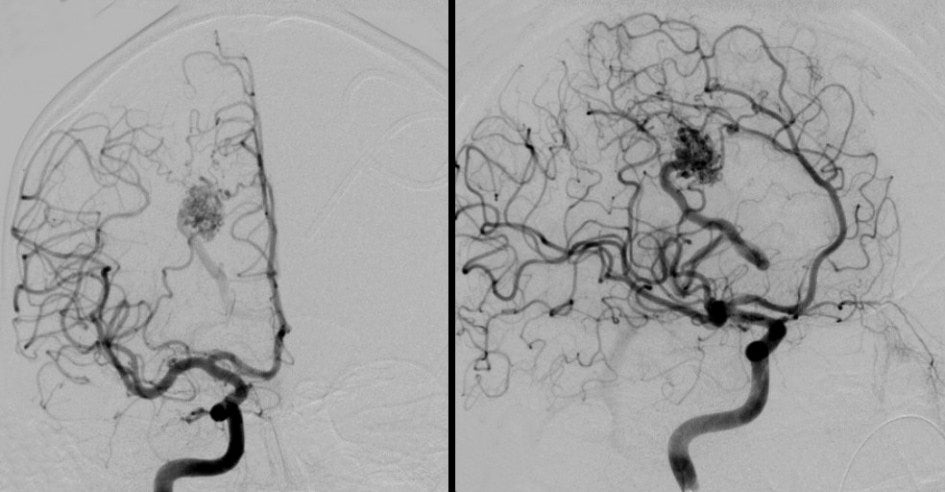
| Arteriovenous Malformation (AVM):
Cerebral angiogram: Right internal carotid artery (ICA) injection. (Left) AP
view; (Right) Oblique view. Note the tangle of blood vessels and the
large draining vein of the AVM. Also note that the arterial feeders
arise from both the middle cerebral artery (MCA) and the anterior
cerebral artery (ACA). To see this patient's MRI,
click here. Arteriovenous malformations (AVMs) are a congenital abnormality of blood vessels. They consist of a tangle of abnormal vessels supplied by arterial feeders and often drained by large dilated veins. AVMs most often occur in isolation. Rarely, they are associated with genetic disorders, among them: Osler-Weber-Rendu syndrome (hereditary hemorrhagic telangiectasia), Sturge-Weber disease, and von Hippel-Lindau syndrome. AVMs are often asymptomatic. Symptoms, when present, may include: • headaches (in some cases a unilateral throbbing headache, mimicking a migraine headache) • seizures (focal, or focal to generalized) • focal neurological deficits • bleeding (may mimic subarachnoid hemorrhage from an aneurysm; bleeding from AVMs account for 2% of all strokes) Larger AVMs are often seen on CT or MRI. Angiography is required to define the vascular anatomy and plan appropriate treatment. Treatment may involve surgical resection, embolization or radiotherapy. |
Revised
11/29/06
Copyrighted 2006. David C Preston