
lens
Detecting Oral Cancer
CWRU researchers devise new approach
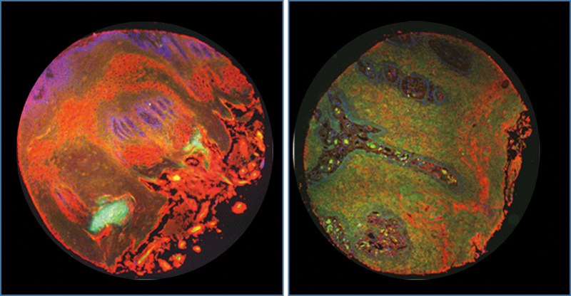 IMAGE: Courtesy of Aaron WeinbergOn the left is healthy tongue tissue stained red by good protein; on the right is cancerous tongue tissue stained green by an over-expression of the protein indicating cancer.
Oral cancers and precancerous mouth lesions are considered especially difficult to diagnose early and accurately.
But a team of researchers, led by Aaron Weinberg, DMD, PhD, chair of biological sciences at Case Western Reserve University School of Dental Medicine, has developed a noninvasive, low-cost test to detect oral cancer, monitor precancerous lesions and determine when a biopsy is warranted. The test involves assessing the levels of two proteins in cells brushed from suspicious oral lesions. A high score implies cancer and indicates the need for a biopsy.
The new patented approach can reduce unnecessary biopsies, Weinberg said, and be particularly beneficial in under-resourced countries where oral cancer is rampant and pathology services are insufficient. The study was published in Cell Reports Medicine.