Department
of Biomedical Engineering
Applied
Neural Control: Prof. J.Thomas Mortimer
Thu. May 02 2024
| Department
of Biomedical Engineering |
|||
| middle |
Applied
Neural Control: Prof. J.Thomas Mortimer |
||
|
Thu. May 02 2024
|
|||
|
|
||||||||||||||||||||||||||||||
| . | . |
|
Updated : August 20, 2014 |
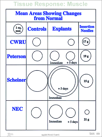 Relative areas showing tissue changes
in the implanted muscles.
Relative areas showing tissue changes
in the implanted muscles.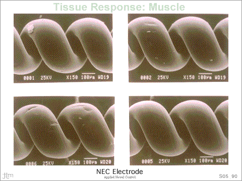 (At Left) Scanning electron micrograph of the insulated coil
section from an as-packed NEC electrode, showing adherent
particulate material on the surface.
(At Left) Scanning electron micrograph of the insulated coil
section from an as-packed NEC electrode, showing adherent
particulate material on the surface.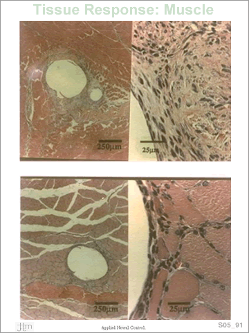 (Top) Tissue from control muscle after 7 days of electrode
implantation; section at level of the barb. Right: magnified
view of tissue between the barb and coil (H & E stain).
(Top) Tissue from control muscle after 7 days of electrode
implantation; section at level of the barb. Right: magnified
view of tissue between the barb and coil (H & E stain).