
Potential biomarker of disease appears to be free of troubling image variability
Preliminary computerized imaging reveals the shape of the prostate and a compartment within the gland—called the transitional zone—consistently differ in men with prostate cancer than those without the disease, according to new research led by Case Western Reserve University. The finding may provide a new avenue to diagnose the disease—perhaps even the cancer’s aggressiveness. The differences held up in comparisons of magnetic resonance imaging (MRI) scans of 70 patients. The scans came from three different medical institutions in Ohio and two in Sydney, Australia, on different makes and models of MRIs. The research is published in Scientific Reports. “Looking at shape is a fundamental shift from looking at the intensity of pixels in an image to predict if a patient has prostate cancer,” said the Anant Madabhushi, F. Alex Nason professor II of biomedical engineering and leader of the research. “Pixel intensities vary, but shape is resilient.” Variability in MRI scans can result in disagreement as to whether prostate cancer is present, in turn potentially resulting in unnecessary biopsies and treatments. The American College of Radiology and others are working to develop standards to eliminate inconsistencies in imaging.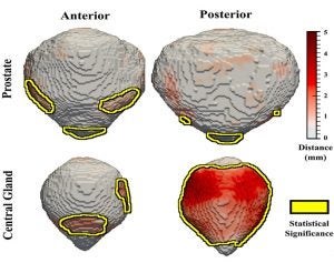 Atlas-based population analysis revealed differences in the shape the prostate and its subregion, the central gland, between normal and malignant prostates. Prostate cancer seems to induce significant changes in the shape of the prostate apex, while benign prostatic hypertrophy appears to drive the differences observed on the posterior side of the central gland for patients without cancer
“Here, we potentially have an image-based biomarker for prostate cancer, which is not greatly sensitive to the MRI parameters used by each institution, the maker of the MRI or the scanner itself, ” Madabhushi said.
Atlas-based population analysis revealed differences in the shape the prostate and its subregion, the central gland, between normal and malignant prostates. Prostate cancer seems to induce significant changes in the shape of the prostate apex, while benign prostatic hypertrophy appears to drive the differences observed on the posterior side of the central gland for patients without cancer
“Here, we potentially have an image-based biomarker for prostate cancer, which is not greatly sensitive to the MRI parameters used by each institution, the maker of the MRI or the scanner itself, ” Madabhushi said.