Hi! I’m a current undergraduate from Maumee, OH majoring in both biology and dance on the pre-medicine track. I’ve always been interested in how the demands of dance impact human biology, specifically the bones. Having experienced several skeletal injuries during my time as a competitive dancer, I was excited to come to CWRU to look at dance using a scientific lens. The craniofacial project in the Atit lab has provided me with a unique opportunity to study orthopedics using a mouse model of craniosynostosis. In fact, the work I have done in the Atit lab has inspired me to pursue further orthopedic research during my time in medical school and beyond.
My Project
Craniosynostosis (CS) is a congenital defect in which the flat bones at the top of the skull fuse too early during development. This results in the loss of one or multiple cranial sutures, which are required for these calvarial bones to adapt to and protect the growing brain. Currently, reconstructive surgery is the only available therapy for this condition. However, it has been noted that dysregulation of the extracellular matrix protein, fibronectin (FN), in the cranial mesenchyme is associated with CS. The goal of my project is to understand this connection at the cellular level, which may eventually lead to therapies that are less invasive and financially draining than surgery.
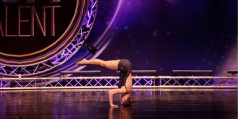
This is me starting a headstand at a dance competition.
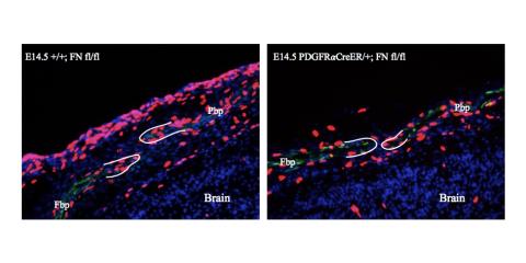
Coronal suture between the developing frontal (Fbp) and parietal bone primordia (Pbp) in transverse sections of E14.5 mouse embryos. Dividing cells are labeled in red.