Projects
These are the projects currently underway in the Basch lab:
Development of the Stria Vascularis
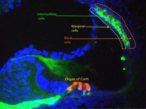
The stria vascularis (SV) is a specialized epithelial structure of the mammalian cochlea that produces endolymph, a potassium-rich fluid responsible for the positive endocochlear potential of the scala media. This positive extracellular potential is the major driving force for proper signal transduction by mechanosensory hair cells and thus normal hearing. The SV is localized in the lateral wall of the cochlea and consists of a multilayered, highly-vascularized epithelium. It is composed of three main cell types with distinct embryonic origins:
- Lining the scala media are the marginal cells derived from the otic epithelium;
- The intermediate cells are specialized melanocyte-like cells derived from migratory neural crest cells and;
- The basal cells are adjacent to the fibrocytes of the spiral ligament and are derived from otic mesenchyme.
Endodermally derived blood vessels are interspersed between these cells throughout the SV.
Defects in the development of the stria vascularis are responsible for a number of inheritable deafness syndromes in humans, including Waardenburg syndrome, Jervell and Lange-Nielsen syndrome, Bartter syndrome and SeSAME syndrome. Strial degeneration is estimated to cause more than 30 percent of age related hearing loss or presbycusis which affects between 25–40 percent of the population over 65 years and over 60 percent of the population over 80 years old.
In spite of its importance for normal hearing, very little is known about how the stria vascularis develops and how or why it degenerates as we age. Our goal is to understand the development of the stria vascularis and apply this knowledge towards therapies for repair and regeneration of the stria in cases of congenital defects and age-related hearing loss.
Development of the intermediate cells of the stria vascularis
Our initial studies of the stria vascularis will focus on the development of intermediate cells. A similar approach will be developed for marginal cells and basal cells. Eventually we will integrate these studies to analyze how all the strial cell types interact in development and degeneration.
Intermediate cells of the stria are derived from neural crest cells. These cells originate in the dorsal neural tube of the embryo and migrate extensively throughout the body. In addition to intermediate cells, neural crest give rise amongst other derivatives to the peripheral nervous system, all of the pigmented cells in the body and most of the craniofacial cartilage and bones.
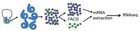
Using Cre-Lox technology, we genetically labeled intermediate cells of the stria vascularis. This allowed us to isolate and sequence mRNA from intermediate cells. We generated a list of genes that are specifically upregulated in these cells at neonatal stages. With this information, we can now begin to ask some questions about the development of these cells:
- When and how do neural crest migrate to the inner ear?
- How do neural crest differentiate into intermediate cells of the stria?
- How do intermediate cells interact with other cell types of the stria?
- Are intermediate cells necessary for proper vascularization of the lateral wall?