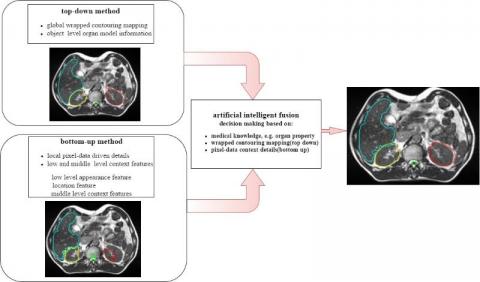
Atallah Baydoun MD and Fang Liang PhD, a Biomedical Engineering PhD student and Senior Research Associate in the Quantitative Imaging Laboratory (QIL), were recognized for work using machine learning methods to automatically draw the boundaries or contours of several organs in MR images. The automated method is faster and more consistent than physicians who currently complete the task in the clinic. This is an important step toward clinical implementation of MR guided adaptive radiation therapy. Organs and tumors change in size, shape, and position over the course of therapy. Recent studies showed that adjusting the contours achieves better dosimetric coverage and may improve survival for some patients undergoing radiation therapy. The QIL is a multi-disciplinary laboratory with physicians, scientists, and engineers bridging radiology, radiation oncology and biomedical engineering at University Hospitals Seidman Cancer Center and Case Western Reserve University School of Medicine. This work was initiated with Dr. Liang and is a collaboration with teams from Jiangnan University in China and Washington University in St. Louis. This is one of three presentations the QIL will be making at this year's RSNA conference in Chicago, the largest imaging conference in the world.
Link to the article at AuntMinnie : AI may enhance MRI-guided adaptive radiation therapy