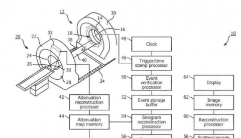
Method for modeling and accounting for cascade gammas in images has been published as US Patent Application 20160048615. Inventors are Professor Raymond Muzic, PhD who is the scientific director of the Quantitative Imaging Laboratory, Dr. Jeffrey Kolthammer, PhD who is a CWRU PhD graduate, and Dr. Jinghan Ye, PhD. The invention occurred in the context of an academic-industrial collaborative project supported by the Ohio Department of Development, “Absolute Myocardial Blood Flow,” Ohio Third Frontier Grant TECH 11-046. CWRU, Philips Healthcare, and University Hospitals Case Medical Center were partners in the project. Other key people were Dr. James O’Donnell, MD who is section chief of UH Nuclear Medicine, Mr. Piotr Maniawski who is Director of Clinical Science, Advanced Molecular Imaging of Philips and Mr. Jeff Kaste who is Director of Collaborations and Grants, Advanced Molecular Imaging of Philips.
The invention improves the quantitative accuracy of the cardiac and other PET images collected using 82Rb. The basis for PET imaging is coincident detection of a pair of gamma rays produced after a positron ejected is in radioactive decay. A complication is that 82Rb is not pure positron emitter; rather additional gamma rays - cascade gammas - are produced during the radioactive decay. When the PET scanner detect a cascade gamma and confuses it with an annihilation gamma ray, the events are mispositioned and there is a loss of quantitative accuracy. By using Monte Carlo simulation, the effect can be modeled and corrected.


