Transformative eye research expands donor pool for corneal transplant patients
Study shows corneas from donors with diabetes are just as effective for surgery
Many eye banks won’t accept corneas from donors with diabetes, concerned they might be harder to prepare for transplant surgery or are more likely to fail.
But a new study led by researchers at Case Western Reserve University and University Hospitals suggests otherwise. The results, published Oct. 17 in the journal JAMA Ophthalmology and presented at the national meetings in Orlando of the American Academy of Ophthalmology, Cornea Society and Eye Bank Association of America, found no significant differences in patients who received corneas from donors with diabetes than from those without the disease one year after the surgery.
The implication is that the number of corneas available for transplant worldwide could be significantly increased.

“We also found the severity of the donor’s diabetes did not affect transplant outcomes, and the corneal structure remained healthy across all groups,” said Jonathan Lass, the Charles I Thomas Professor in the Department of Ophthalmology and Visual Sciences at the Case Western Reserve University School of Medicine and the study’s lead researcher.
The study
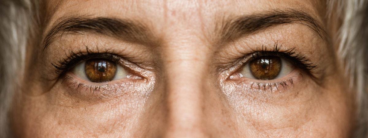
Like a camera lens, the cornea is the clear, dome-shaped window in front of your eye that helps focus light. Vision can occasionally be blocked or distorted by the cornea becoming cloudy, scarred, swollen or misshaped—often caused by complications from illness, injury or surgery.
In the study, funded by the National Eye Institute (NEI) of the National Institutes of Health, 1,097 patients had what is known as Descemet Membrane Endothelial Keratoplasty (DMEK), transplant surgery that replaces the innermost, diseased layer, the endothelium, a single layer of cells that lines the back of the cornea and helps keep the cornea clear.
Two-thirds of patients received corneas from donors without diabetes and one-third from donors with the disease. After one year, there was no difference in transplant success between the two groups.
The study involved donated corneas from 13 eye banks in the United States, 28 clinical testing sites and 46 surgeons. The patients averaged 70 years old.
“The DEKS findings are good news for eye banks because they demonstrate that people with diabetes can be cornea donors, increasing the number of people whose sight can be restored, and lives transformed, through transplant procedures,” said Kevin Corcoran, president and CEO of the Eye Bank Association of America.
Fate of vital transplanted cells
A concurrent related study tracked what happens to the corneal endothelial cells after the procedure. These cells are important to pump fluid out of the cornea to keep it clear. Unlike other cells in the body, endothelial cells do not regenerate. Once they are lost, they can’t be replaced naturally.
Using a special microscope, these cells can be photographed without touching the eye. The images can then be analyzed over time to determine changes in the number, size and shape of the cells. This image analysis work was done by the Cornea Image Analysis Reading Center at Case Western Reserve and University Hospitals Eye Institute, led by professor and Director Beth Ann Benetz.
“We found that that endothelial cell loss one year after surgery was 28%, regardless of whether the donor had diabetes or not,” Lass said. “These results support no restrictions on the use of tissue from donors with diabetes for DMEK and thus expand the donor pool for corneas.”
“The findings from both studies provide reassuring evidence that corneas from donors with diabetes perform just as well as those from non-diabetic donors,” said Barry Lee, president of the Cornea Society, an international non-profit scientific society that promotes the exchange of information about the cornea and external eye disease.
The NEI has agreed to support studying the participants five years after their surgery to see if the results hold up.


