CWRU researchers’ out-of-the-box investigation on skull bone growth and fusing processes
Radhika P. Atit, PhD
Professor in the Department of Biology at the College of Arts and Sciences
The inspiration for out-of-the-box research ideas might sometimes come from “dreams.”
That was the case for the latest research of Radhika P. Atit, PhD, professor in the Department of Biology at the College of Arts and Sciences who sought to better understand how the mammalian skull grows and fuses, especially before birth.

An infant’s braincase—the roof of the skull that covers the brain—is not a unified entity. It is made up of eight flat bones, connected by flexible joints called sutures. This flexibility enables the skull to accommodate the infant brain as it continues to grow.
Defects occur when one or more sutures close prematurely, leading to a condition called craniosynostosis—the second most common craniofacial birth defect.
Since the fused suture restricts brain growth, the expanding brain pushes the skull to grow abnormally, resulting in an unusual head shape. Yet, the mechanism by which skull bones grow faithfully to encase the brain across vertebrates remained largely unknown to scientists—until recent investigations by Atit’s team and her collaborators.
As Atit pondered the research question in mouse embryos, she began to dream about Spider-Man, in which the skull bone cells “crawled” along Spider-Man’s silk and “met” at the top of the head. This dream, interestingly, aligned with Atit’s unconventional hypothesis that skull bones grow by cells traveling and laying down new tissue as they move.
“At the start of our project, there was no direct evidence showing that the skull bones would grow by physical cell migration; no other bone does that in your body,” Atit said. “If I told somebody that the bone has grown by cells physically moving in space, they would find it unbelievable.”
Still, the image lingered in Atit's mind. Knowing how rapidly the skull grows during mouse embryonic development, she thought that the growing-by-moving hypothesis might not be as implausible as it first seemed.
“In developmental biology, if you have a theory on how a developmental process works, the best way to truly test it is to perturb the system and see what breaks,” Atit said.
Atit then teamed up with Matthew Harris, PhD, professor of genetics and orthopedic surgery at Boston Children’s Hospital and Harvard Medical School. Together, they assessed whether preventing the skull bone cells from moving would interfere with their growth.
Harris, an expert geneticist, used a genetic tool to remove the skull bone cells' ability to “crawl” and migrate in the mouse embryo. The experiment showed that these immobilized skull bone cells could not spread across the skull surface, resulting in small, underdeveloped bones.
“To me, that was the best evidence to date demonstrating that this movement machinery is required for the skull bones to grow and cover the head,” Atit said.
The collaboration was awarded a five-year, $3.2 million R01 grant from the National Institute of Dental and Craniofacial Research at the National Institutes of Health to support their investigation.
The “road” where skull bone cells move on
Atit’s team also considers other factors that may affect the movement of bone cells.
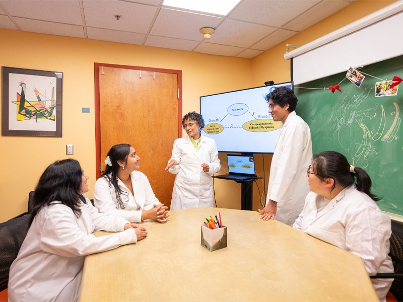
In collaboration with cell movement expert Sevan Hopyan, MD, PhD, of Sick Kids Hospital and the University of Toronto, the scientists investigated the “road” or the “silk” along which the bone cells “crawl”. They hypothesized that fibronectin—a protein outside the cell that supports tissue structure—is one of the “road elements” that is a key support for skull bone growth.
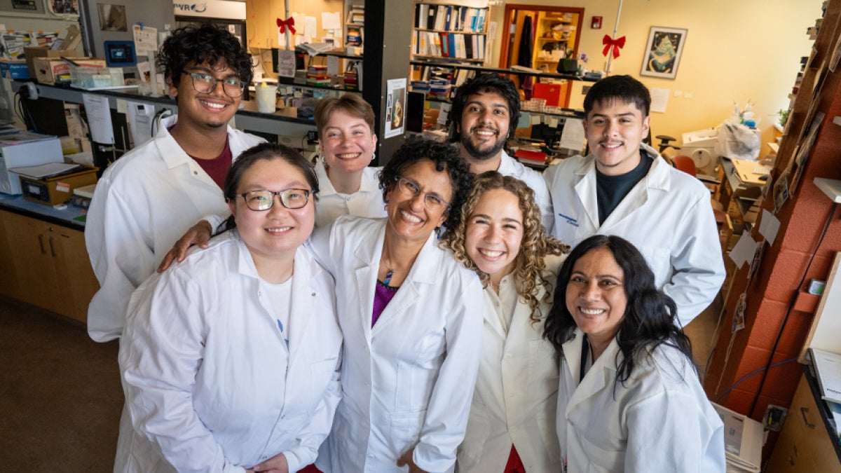
Working with Greg Holmes, PhD, an expert on craniosynostosis at Icahn School of Medicine at Mount Sinai, Atit and her student researchers—Xiaotian Feng, PhD (GRS '24), Megan Gregory, (CWR '24) Alex Flores (CWR '25), Isha Gupta (CWR '24), and Morgan Horowitz, a fourth-year student studying biology and communication sciences—looked into various mouse models of craniosynostosis, whether as a stand-alone disease or a syndromic disease (accompanied by other symptoms). The result is surprising but consistent: abnormal fibronectin “road,” in either their quantity or expression pattern, across six models.
Some of their findings were published in the journal Development, while additional results are currently under peer review.
To date, skull bone defects are most commonly treated with repeated surgeries. The understanding of fibronectin’s significance, as well as the skull bone mechanism of growth, may open up new directions for less invasive treatment.
“I only want to work on problems that keep me up at night,” Atit said. “They have to capture my imagination. And I want to pursue problems that inspire others to think differently and ultimately transform discoveries into faster cures.”