|
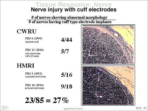
 Typically, investigators have reported no outward signs
of nerve trauma. The concern is how to interpret these findings
in relationship to safety of cuff type electrodes. Typically, investigators have reported no outward signs
of nerve trauma. The concern is how to interpret these findings
in relationship to safety of cuff type electrodes.
In the upper right panel is shown an image of a typical “crescent
type region” It is characterized by having numerous
thinly myelinated large axons, a loose axon packing density,
interneural fibrous tissue, and subperineual thickening.
Macrophages do not seem to be present in the “crescent
type regions”. These “crescent type regions” are
characteristically crescent in shape and positioned on a
superficial region of the fascicle. In the panel on the lower
right is a cross-section from a “normal” fascicle.
The most common type of mechanically induced injury is manifested
as subperineurial crescents of endoneurial connective tissue.
These areas of increased connective tissue were most frequently
situated at the periphery of the fascicles and occasionally
were accompanied by degenerating axons, localized axonal
loss, and remyelinating axon profiles [8]. The subperineurial
crescents may be adaptive to some degree since they closely
resemble ‘Renaut bodies’ (hyaline-appearing,
loosely-textured, whorled, cell-sparse structures found in
the subperineurial space) [13]. Weis et al. [14] reported
an association between the development of Renaut bodies and
nerve entrapment, and proposed that these structures may
serve as a pressure-absorbing cushion for entrapped nerve.
Agnew, W.F., and D.B. McCreery. "Principles of safe
and effective nerve stimulation", In: New perspectives
in sacral nerve stimulation, edited by U. Jonas, and V. Grunewald.
London: Martin Dunitz, 2002, pp. 29-42
|

