GARLA (Gross Anatomy, Radiology, Living Anatomy) is one of the components of the Structure block that stretches longitudinally across the first two years of medical education at Case Western Reserve University School of Medicine.
Structure integrates basic and clinical concepts of GARLA and Histopathology. An understanding of these disciplines forms a framework for understanding the basic mechanisms that underlie health and disease. The overall learning objectives of this longitudinal block are to develop an understanding of macro-, micro-, and ultramicroscopic human structure, nomenclature, imaging techniques, basic physical examination skills related to these structures, and their respective functions in normal and diseased organs, tissues, and cells. We attempt to provide opportunities for students to apply and view the material we cover as they would in a clinical setting.
The knowledge of normal gross and microscopic anatomy, as well as imaging (radiology) of these organs and tissues, is necessary for an appreciation of the relationships between altered structure and disturbed function as well as a foundation to understand the anatomical basics of physical examination skills. Thus, Structure bridges the normal and diseased, and begins to prepare students for the transition from classroom to ward.
GARLA begins in the first block titled, "Becoming a Doctor", with a peer-led introduction to anatomy and radiology. Second-year students meet weekly with small groups of first-year students and discuss and discover the foundations of the basic concepts underlying these disciplines.
Intervening between the first two blocks is a two-week dissection Boot Camp. During Boot Camp, groups of four students dissect one body cavity (either thorax or abdomen) and one limb (either upper or lower limb) and present that material to their peers who dissected the other region. There are formative and summative assessments at the end of weeks 1 and 2 respectively. The Boot Camp culminates with a body donor memorial service where students and donor families gather to recognize the contributions of our donors.
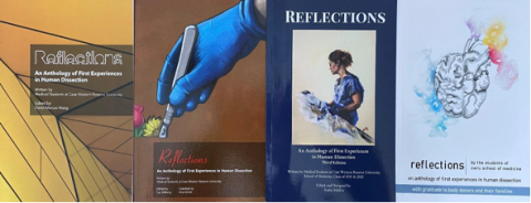
After their first dissection, students are asked to write a reflection about their “first cut” experience, and approximately every second year a Reflections an internal, peer reviewed book is published by the medical students. These books are distributed to the donor families at the memorial service.
3-part GARLA Curriculum
After the Boot Camp, the students begin the 3-part GARLA curriculum which is integrated
Virtual Anatomy Lab
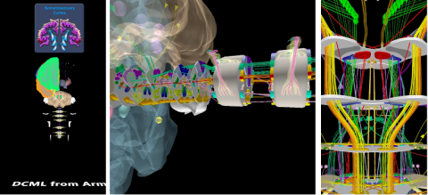
In the virtual anatomy lab, students use HoloAnatomy to review the anatomy and neuroanatomy associated with the theme of the day. HoloAnatomy is a mixed reality anatomy application created by a team of anatomists, artists, computer programmers and instructional designers at Case Western Reserve University. It includes a library of digital 3-D Anatomy Art with of over 9,000 anatomical asserts. Each lesson is custom-made to focus on the day’s topic. HoloAnatomy expands upon our cadaver-based teaching practices.
Our research has found that our medical students learn as well with HoloAnatomy as they do in the cadaver lab but with less time invested. This has enabled us to integrate equal amounts of radiology and physical exam and ultrasound in every GARLA session.
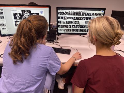
GARLA Radiology Reading Room
In the GARLA radiology reading room, pairs of students scroll through relevant images and learn to interpret them. The focus is on normal anatomy and appropriate tests to order in association with the day’s anatomical theme.
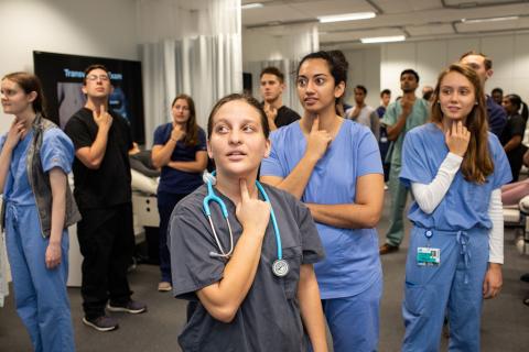
The Living Anatomy Room
In the Living Anatomy room, groups of four students explore the normal anatomy associated with the day’s theme as they examine standardized patients using physical exam skills as well as cart-based and hand-held ultrasound devices.

We believe that through the GARLA curriculum we enable our students to apply the anatomy they are learning to bring anatomy to life.
Relevant Bibliography
Zuniga, B. and Wish-Baratz, S. (2015). Anatomy Dissection Guide – A Comparative Study of Formats. Med Sci Educ. 25 (3), 271. doi:10.1007/s40670-015-0135-5.
Wish-Baratz, S., Gubatina, A. P. Jr., Enterline, R., and Griswold, M.A. (2019) A New Supplement to Gross Anatomy Dissection: HoloAnatomy. Med Educ, 53, 522. doi: 10.1111/medu.13845
Baratz, G., Wilson-Delfosse, A.L., Singelyn, B.M. Allan, K.C. Rieth, G.E., Ratnaparkhi, R., Jenks, B.P., Carlton, C., Freeman B.K. and Wish-Baratz S. (2019). Evaluating the Anatomage Table Compared to Cadaveric Dissection as a Learning Modality for Gross Anatomy. Med Sci Educ. 29(2), 499. doi: 10.1007/s40670-019-00719-z.
Stojanovska M., Tingle G., Tan L., Ulrey L., Simonson-Shick S., Mlakar J., Eastman H., Gotschall R., Boscia A., Enterline, R., Henninger E., Herrmann KA., Simpson S.W., Griswold MA., Wish-Baratz S. (2020). Mixed Reality Anatomy using Microsoft HoloLens and Cadaveric Dissection: A Comparative Effectiveness Study. Med.Sci.Educ. 30, 173–178. doi: 10.1007/s40670-019-00834-x.
Ruthberg, J., Tingle, G., Tan, L., Ulrey, L., Simonson-Shick, S., Enterline, R., Mlakar, J., Eastman, H., Gotschall, R., Henninger, E., Griswold, MA., Wish-Baratz, S. (2020). Mixed reality as a time-efficient alternative to cadaveric dissection. Med Teach.42(8), 896-901. doi: 10.1080/0142159X.2020.1762032.
Wish-Baratz, S., Crofton, A.R., Gutierrez, J., Henninger, E., Griswold, M.A. (2020). “Assessment of Mixed Reality Technology Use in Remote Online Anatomy Education.” JAMA Netw. Open. 3(9):e2016271. doi:10.1001/jamanetworkopen.2020.16271.
Wish-Baratz, S., Jones, B., Navracruz, L.C., Rowland-Seymour, A., Herrmann, K.A. (2020). “GARLA: Integrating Traditional and Modern Methodologies in Anatomy Education.” Med Sci Educ. 30(4), 1727-1728. doi: 10.1007/s40670-020-01067-z.
Deming, N., Singer, M., Baratz, G., Wish-Baratz, S. (2021). Matriculating Students’ Opinions on Cadaveric Dissection: Maintaining Tradition in Changing Times". Med Sci Educ. 31(1), 41-44. doi: 10.1007/s40670-020-01139-0.
Thom, M.L., Kimble, B.A., Qua, K., Wish-Baratz, S. (2021). “Is remote near-peer anatomy teaching an effective teaching strategy? Lessons learned from the transition to online learning during the COVID-19 pandemic.” Anat Sci Educ.14(5), 552-561. doi: 10 DOI:10.1002/ASE2122.
Baratz, G., Sridharan, P.S., Yong, V., Tatsuoka, C., Griswold, M.A., Wish-Baratz, S. (2022). “Comparing Learning Retention in Medical Students using Mixed-Reality to Supplement Dissection: A Preliminary Study. Int J Med Educ. 13, 107-114. doi: 10.5116/ijme.6250.0af8.