Department
of Biomedical Engineering
Applied
Neural Control: J.T. Mortimer
Tue. Jul 21 2026
| Department
of Biomedical Engineering |
|||
| middle |
Applied
Neural Control: J.T. Mortimer |
||
|
Tue. Jul 21 2026
|
|||
|
|
||||||||||||||||||||||||||||||
| . | . |
|
Updated : August 20, 2014 |
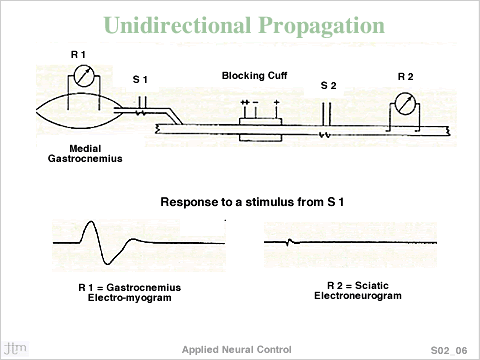 The
experimental preparation is shown in the top of the Figure
was designed to test the hypothesis that unidirectionally
propagating action potentials were actually generated. R1
and R2 are electro-myogram and electro- neurogram recording
sites respectively. S1 and S2 are hook electrodes used to
apply currents to the nerve trunk for test stimuli. The tripolar
nerve cuff is in the center, around the nerve trunk. Action
Potentials (APs) generated under the central contact ('cathode')
are arrested at the contact on the left, which carries more
current than the one on the right and allowed to propagate
to the right, because the contact on the right carries less
current than the one on the left but just enough current
to suppress the development of a virtual cathode on the left
side.
The
experimental preparation is shown in the top of the Figure
was designed to test the hypothesis that unidirectionally
propagating action potentials were actually generated. R1
and R2 are electro-myogram and electro- neurogram recording
sites respectively. S1 and S2 are hook electrodes used to
apply currents to the nerve trunk for test stimuli. The tripolar
nerve cuff is in the center, around the nerve trunk. Action
Potentials (APs) generated under the central contact ('cathode')
are arrested at the contact on the left, which carries more
current than the one on the right and allowed to propagate
to the right, because the contact on the right carries less
current than the one on the left but just enough current
to suppress the development of a virtual cathode on the left
side.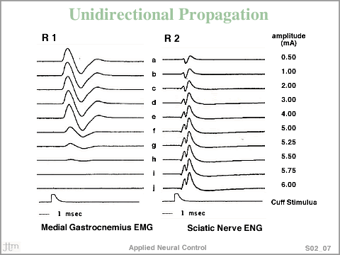 Above
a certain current amplitude, the cuff electrode causes arrest of APs towards the muscle (left) while allowing them to escape to the right.
Above
a certain current amplitude, the cuff electrode causes arrest of APs towards the muscle (left) while allowing them to escape to the right.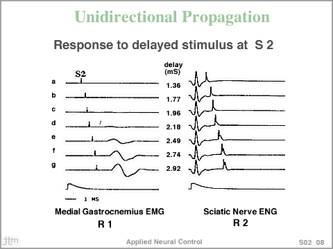 If
unidirectionally propagating (moving to the right) were actually
generated at the cuff electrode, the nerve fibers carrying
these APs would be refractory for a period of time after
the APs had passed the region of the S2 electrode. The experimental
data shown in the figure support the hypothesis. Stimuli
from the tripolar electrode 'arrests' APs propagating towards
the Gastrocnemius, while allowing them to escape antidromically.
Only when the S2 stimuli were applied at delays greater than
2.4 msec was the S2 stimulus capable of exciting the nerve
to cause muscle contraction. This implies that a unidirctionally
propagating pulse was generated and the refractory period
was greater than 2.18 msec.
If
unidirectionally propagating (moving to the right) were actually
generated at the cuff electrode, the nerve fibers carrying
these APs would be refractory for a period of time after
the APs had passed the region of the S2 electrode. The experimental
data shown in the figure support the hypothesis. Stimuli
from the tripolar electrode 'arrests' APs propagating towards
the Gastrocnemius, while allowing them to escape antidromically.
Only when the S2 stimuli were applied at delays greater than
2.4 msec was the S2 stimulus capable of exciting the nerve
to cause muscle contraction. This implies that a unidirctionally
propagating pulse was generated and the refractory period
was greater than 2.18 msec.