The first XFP endstation, termed the High-Dose Endstation, is centered at 28m from the source, at the 1:1 focus of the beamline’s front-end toroidal focusing mirror. At this location, beam sizes as small as 100 μm x 400 μm (vert x horz., FWHM) can be achieved, permitting very high flux densities and beam powers to be delivered to the sample (photon flux ~1016 photons/sec, maximum power of 500 W/mm2 at 500mA NSLS-II ring current). This endstation is well-suited for X-ray footprinting of highly scavenging systems requiring an intense hydroxyl radical pulse, such as membrane proteins, large multi-component protein complexes, and investigations under in vivo conditions. Most experiments are conducted by flowing samples through Kapton-coated glass capillaries (200 μm and 530 μm IDs standard), with the cross-section of the beam and the flow rate defining X-ray exposure times.
The centerpiece of this endstation is a flexible three-axis motorized table that can support a wide variety of apparatus (Figure 1). Standard configurations available for user experiments include a continuous flow pumping scheme with incubator and fraction collector for in vivo footprinting (Figure 2), and a capillary flow setup using a syringe pump for sample delivery (Figure 3) for equilibrium measurements at high X-ray/radical doses. Non-standard endstation setups provided by users can also be accommodated at this location, following discussion with beamline staff.
For details of the performance of XFP at the high-dose endstation, see our recent beamline paper (Asuru et al., J. Synch. Rad. 2019, 26, 1388-1399. DOI: 10.1107/S1600577519003576). Protocols for performing in vivo footprinting experiments can be found in a recent article from Sarah Woodson’s group at Johns Hopkins University (Hao et al., Current Protocols in Nucleic Acid Chemistry, 2018, e52. DOI: 10.1002/cpnc.52).
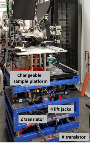
Figure 1. Three axis motorized experimental table for the High-Dose Endstation. This flexible table is equipped with a 36 in. square breadboard and can handle several hundred lbs of equipment. It features a changeable sample platform, 4 lift jacks, a Z translator, and an X translator.
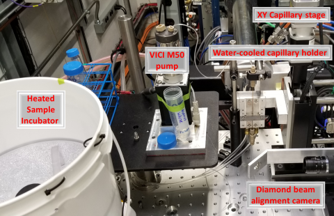
Figure 2. In vivo footprinting setup. The VICI M50 continuous flow pump moves sample from the heated sample incubator through a glass capillary intersecting X-ray beam in the water-cooled capillary holder, and then to a fraction collector (hidden). The capillary can be precisely aligned into the beam using a motorized XY stage and a diamond screen based beam alignment camera.
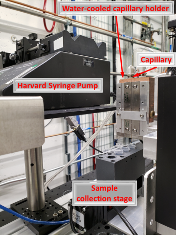
Figure 3. Conventional capillary flow setup with water-cooled capillary holder using a Harvard syringe pump for sample delivery and a small sample collection stage for exposed samples. Several capillary diameters are available; most experiments are performed using a 200 μm ID capillary permitting exposure times of 75 μs to 1 ms.
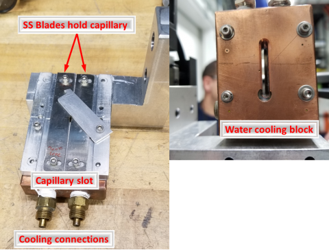
Figure 4. Close-up view of the water-cooled capillary holder. The capillary is held between 2 stainless steel blades in thermal contact with a water-cooled copper block. The assembly is mounted to a translation stage that allows different regions of the capillary to be exposed, reducing damage caused by the X-ray beam.