Affiliated School or College: School of Medicine
The Light Microscopy Imaging Core facilities (LMIC) at Case Western Reserve University provide advanced imaging systems. including whole slide scanning, confocal microscopy, STED microscopy, lightsheet microscopy, and spatial omics. Supported by sophisticated data analysis software, these systems enable detailed studies of cells, tissues, and organisms, driving significant research discoveries. Join us in advancing scientific explorations!
Learn more about the Light Microscopy Imaging Core
Our instrument portfolio
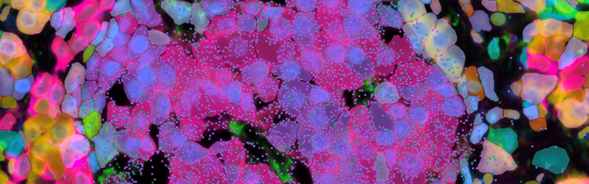
Imaging-Based Spatial-Omic Platforms
- Xenium — 10x Genomics
- CosMx/AtoMx — Bruker Spatial Biology
LightSheet Microscopy
- Cleared-Tissue LightSheet - 3i
- Lattice LightSheet - 3i
Confocal Microscopy
- HyVolution SP8 - Leica
- TCS SP8 gated STED 3X - Leica
- SPIN SR-10 Spinning Disc - Olympus
Whole Slide Scanner
- NanoZoomer S60 (Brightfield and Fluorescent slides) — Hamamatsu Photonics
Widefield Microscopy
- DM6000 Upright Microscope - Leica
- DM6000 Inverted Microscope - Leica
Analysis Workstation and Remote Data Storage
- Lambda Vector (2 GPUs for deep learning)
- Analysis software
- 250 TB of storage
Customers: External and Internal
Hours
9 a.m. to 5 p.m. on weekdays
Location
Case Western Reserve University
School of Medicine
Robbins Building E632
2210 Circle Drive
Cleveland OH 44106