The Case Western Reserve University School of Medicine Graduate and Medical Student Research Day will take place on Thursday, Oct. 15, from 8:00 a.m. to 4:30 p.m. in the Tinkham Veale University Center. The day will include a keynote address, posters, oral presentations, and awards.
If you have any questions about the event, the abstract submission process, or judging, please contact som_student_research_day@case.edu. We look forward to seeing you at Research Day 2026!
Deadline for Submission
The deadline for abstract submissions is Aug. 31 by 11:59 p.m. The submission site will accept abstracts for posters and oral presentations to be presented on Thursday, Oct. 15, at Tinkham Veale University Center
Abstract and Presenter Eligibility
The criteria below applies to both posters and oral presentations. To submit an abstract and have the opportunity to present, you must be:
- A CWRU SOM graduate or medical student who has conducted research as a student in a CWRU SOM graduate or medical program
- Includes students pursuing MDs, PhDs, and Master’s degrees
- First year students are not generally eligible to present, unless they were formerly in another CWRU School of Medicine graduate program or are in the CCLCM program.
- The primary/presenting author on only one abstract submission for this event each year.
- You may be a co-author on multiple abstracts, but only the primary author will present if selected for an oral presentation, and only the primary author will be recognized with any awards.
You may submit an abstract:
- For work already presented at another meeting or already published, if the work was conducted during your time as a graduate or medical student at the CWRU School of Medicine.
- For work that does not have results at the time of submission but is expected to have results by the time of Research Day for consideration as a poster. These will not be accepted for oral presentations.
- In any field (basic science, clinical/translational research, health services, medical education research, policy-oriented analysis, etc.) using any methodology (quantitative or qualitative).
Additional Abstract Eligibility Applicable to Only Oral Presentations
In order for abstracts to be considered for an oral presentation you must:
- Have results by the time of submission
- Include a link to a video presentation about your abstract within the word document upload portion of this form.
- The required video presentation follows Three Minute Thesis (3MT) format.
- You will have three minutes and one slide to effectively explain your research and its significance in a language appropriate to a non-specialist audience.
- Your video link must be publicly available and included on your abstract document.
- Please watch this 2024 3MT informational session recording from the linked start time until 11:46 for more information about video presentation parameters.
- Judging criteria is also discussed in this clip; remember you are trying to demonstrate both the value of your research and your skill as a presenter to be selected to give a full length (10-12 min) oral presentation.
- The required video presentation follows Three Minute Thesis (3MT) format.
- Additional tips about preparing this type of presentation including designing your slide and generating a link is available in this clip from the same session.
Abstract Preparation Guidelines
Please use the following abstract formatting guidelines:
Heading section (excluded from word count maximum) consists of:
- abstract title
- your name and program
- mentor's name and affiliation
- any other authors you would like to include/recognize
- any acknowledgements (including for funding)
- Consideration for oral presentation (optional): provide LINK TO YOUR VIDEO if you wish to be considered for oral presentation or leave blank if not applicable
Structured abstract with four sections (300 words maximum):
- Significance: (including the research question)
- Methods
- Results: If you do not have results by the abstract deadline, you may still submit. Please include this section and a statement of whether you are likely to have results by the presentation date. Abstracts without results are often accepted for posters but will not be considered for oral presentations)
- Conclusions: Include a statement of the potential significance of the project and next steps for research
Formatting guidelines:
- Maximum of 300 words, Arial font, size 11, single spaced
- Do not insert any tables or figures
- A format template is available for download here.
- An example of an appropriately formatted abstract on a clinical topic is here.
- An example of an appropriately formatted abstract on a basic science topic is here.
Attendance and Presenting Your Work
Attendance
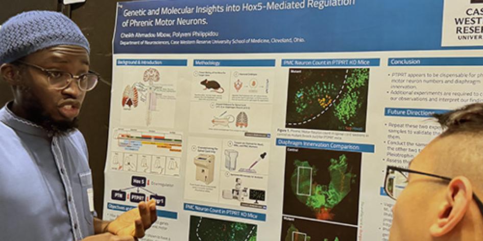
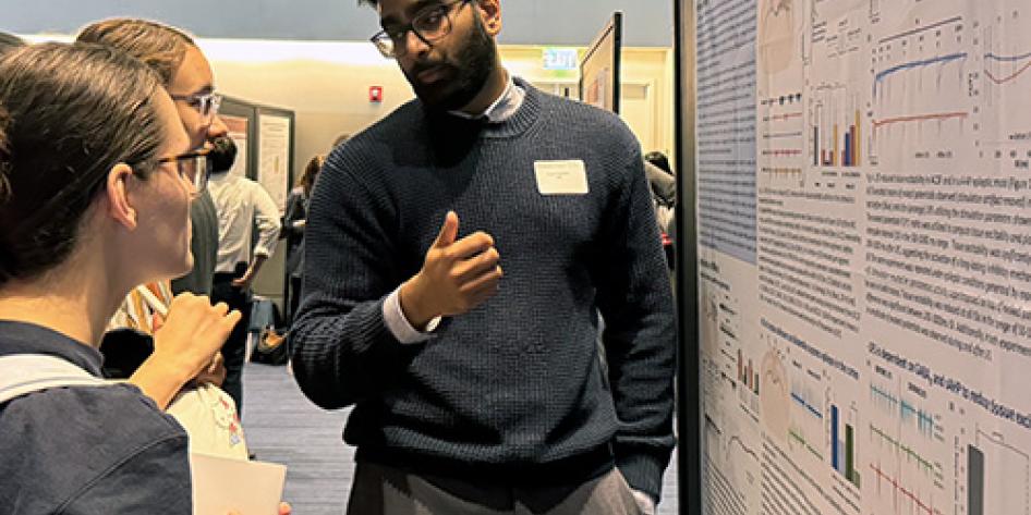
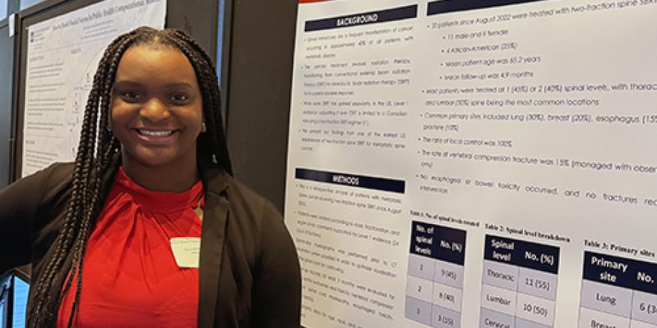
Posters will be scheduled for either the morning (8 am -11:30 pm) or afternoon (11:30 am - 4:30 pm) session of SOM Graduate and Medical Student Research Day on Oct. 15. Sessions will be assigned with a balance of topics and programs in each session.
You are strongly encouraged to attend the entire day, but at a minimum are expected to attend the entire session to which you are assigned, including poster session and Dean's welcome and keynote address (morning), or poster session, oral presentations and awards ceremony (afternoon). Please hold the entire day open on your calendar.
Oral presentations will be scheduled for the afternoon session on Thursday, Oct. 15.
Poster Printing
Please do not print your posters until you receive an acceptance email (by Sept. 21) to confirm if you have been selected for an oral presentation or poster, and which session you have been assigned to. Printing guidelines and suggested printing locations will be shared with you at that time.
Non-presenters
Students who are not presenting are encouraged to attend to learn about their colleagues’ research activities. Everyone is invited to attend; those who are not presenting will be asked to RSVP separately through Campus Groups.
Who is eligible to be a judge?
The CWRU School of Medicine invites faculty, alumni, and senior trainees (residents, fellows, post-docs) to participate in providing student feedback and award consideration.
This judge sign-up site accepts from faculty, alumni and senior trainees who wish to serve as judges:
- For student abstracts; to select those presented as posters or oral presentations during Research Day. Abstract judging takes place via an online form between Sept. 8 and 16.
- For student poster presentations conducted in person 10–11:30 a.m. on Thursday, Oct. 15, at the Tinkham Veale University Center.
- For student poster presentations conducted in-person 1:30–3 p.m. on Thursday, Oct. 15, at the Tinkham Veale University Center.
What is the deadline to sign up to be a judge?
The deadline to sign up to judge via this site is Aug. 31 by 11:59 p.m.
If you cannot help with judging, you are still welcome and encouraged to attend; RSVPs will be requested separately!
Judging Details
Abstract judging:
Abstract judges will be assigned approximately 15 abstracts to evaluate. Some abstracts will also include a linked, 3-minute video presentation to be considered for oral presentations.
Abstract judges will be asked to review both the written abstract and the video presentation, if relevant. Materials will be available no later than Sept. 8, and evaluations must be submitted through an online form by Sept. 16. Faculty receive 1.5 hours of med school teaching credit for every 4 abstracts evaluated. All judges can add this activity to their CV/service report.
Poster judging:
Poster judges will be assigned 4-6 posters to evaluate during a 90-minute session on SOM Graduate and Medical Student Research Day, Oct. 15. Evaluation is done electronically with a simple rubric.
Judges can indicate availability to judge in the morning (10–11:30 a.m.) and/or afternoon (1:30–3 p.m.) session. Judging determines the poster award winners from each session. Faculty receive 1.5 hours of med school teaching credit for every 4 posters evaluated. All judges can add this activity to their CV/service report. Judges will receive free parking at the Tinkham Veale University Center on the day of the event.
Judges can sign up for an abstract and/or poster judging.
We will make every attempt to match judges with abstracts/posters in their area of expertise but cannot guarantee perfect matches.
Students who are not presenting are encouraged to attend to learn about their colleagues’ research activities. Staff and faculty who are not judging are encouraged to attend to support our students. Everyone is invited to attend; those who are not presenting or judging will be asked to RSVP separately through Campus Groups.